Nuclear Medicine Case Study
Background
When scientists first discovered that atoms contain an incredible amount of energy stored in their nucleus, they weren't thinking about electricity or weapons, but how to use it in the treatment of illness. Marie Curie and her husband, Paul Germain, did not patent radium, but published their results in open journals. Shortly after her discovery, radium was everywhere and is still used in almost every major hospital today. Anger's scintillation cameras are used in a wide variety of nuclear medicine applications, including cardiovascular and pulmonary angiography. They use a series of crystals with varying energy levels to produce an image. A single photon interacts with one crystal, while a double count will interact with a different crystal. The energy of a photon depends on its frequency, and the frequency of the photon is determined by the amount of energy that it carries. The higher the energy of the photon, the more accurate the image produced by the camera will be. Anger's scintillation gamma camera was developed in the 1950s, and it remains the main imaging tool in nuclear medicine. Although several designs have emerged since Anger's invention, it is the Anger camera that dominates the field today. Its versatility, cost effectiveness, and performance make it one of the most widely used gamma cameras.
The Bohr-Posner relationship explains how the phosphorus-based tracer in nuclear medicine works. The study of phosphorus metabolism in rat skeletons was revolutionary. It pioneered the idea of dynamic equilibrium and the use of artificial radioactivity. However, there was a delay before the use of phosphorus was used in scintigraphy. Using the International [1]Congress of Radiology and Electricity in Brussels, the Radium Standards Committee met. Marie Curie and Ernest Rutherford were the most influential members of this committee. During the first meeting, Rutherford warned Meyer that Curie wanted the term "curie" to be defined in terms of one gram of radium. The Committee also had to come up with a naming convention for the radioactive particles.
Procedure
Nuclear medicine uses radioisotopes and gamma rays to diagnose health problems. The process of nuclear medicine involves preparing a tracer that is swallowed, injected into a vein, or inhaled as gas. When the tracer enters the body part being studied, it sends out gamma rays that are picked up by a special camera. A computer processes these signals and converts them into two-dimensional pictures. A radiologist or nuclear medicine doctor interprets the pictures.
A radioactive substance is a type of molecule used in nuclear medicine. They emit radiation when they are inside the body. They are useful for treating cancer, tissue overgrowth, and hyperthyroidism. These chemical compounds are given intravenously, ingested, or breathed in. Scanners detect the localized radioactive substance by detecting its presence with scintillation cameras. The data from the detector is then processed in a computer to produce a two or three-dimensional image.
Unlike CT and MRI scans, PET scans are non-invasive and painless, and use small amounts of radioactive material to visualize the body's tissues. PET scans are used in the diagnosis of many conditions, including cancer, brain disease, and cardiovascular disease. During a PET scan, a patient lies on a table in a “PET scanner” that has a “ring of detectors” surrounding them. While CT scans are like PET scanners, a nuclear medicine scientist will be the one to perform the procedure.
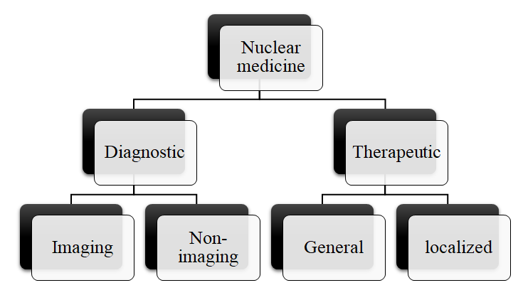
“Types of nuclear medicine”
Applications
Several medical professionals use nuclear medicine to help diagnose and treat a variety of diseases. This type of radiology involves using radiation to generate images that show the health of different parts of the body. The images are digitally generated and transferred to a nuclear medicine physician, who interprets them. Radioactive tracers are usually injected into the vein,
but sometimes can be given by mouth. Typical nuclear medicine scans involve low radiation levels. These tests are also used to detect bone problems, kidney disease, and scars.
Using radioactive materials in medicine has increased substantially over the past decade. Today, it represents one of the largest sources of exposure to the American population, but the extent of this exposure is difficult to determine. The effective dose is a useful measure of potential detriment from exposure to ionizing radiation. Besides its diagnostic value, effective doses also serve as a parameter for determining the appropriateness of examinations involving ionizing radiation.
There are several procedures in nuclear medicine. These procedures can be done in the office or in the hospital. They can be performed for a variety of reasons. For instance, a patient may need to have an MRI of the extremities or of a specific location. These exams take anywhere from 15 minutes to an hour and are relatively painless. Patients can go in for these exams with or without preparation. The patient will need to stay still during the procedure. (Blankenberg 2002)
Other uses of radioisotopes in nuclear medicine include therapy and diagnostic imaging. The irradiation of a tumor can destroy malfunctioning cells. This process is called brachytherapy and is less common than diagnostic use of radioactive material in medicine. The ideal radioisotope is beta emitting, with just enough gamma to allow imaging. When used for cancer therapy, it can be localized in a specific organ or attached to a biological compound....
originally done case solution."}" data-sheets-userformat="{"2":14913,"3":{"1":0},"9":0,"12":0,"14":{"1":2,"2":3355443},"15":"\"open sans\", Arial, sans-serif","16":10}">This is just a sample partial case solution. Please place the order on the website to order your own originally done case solution.

